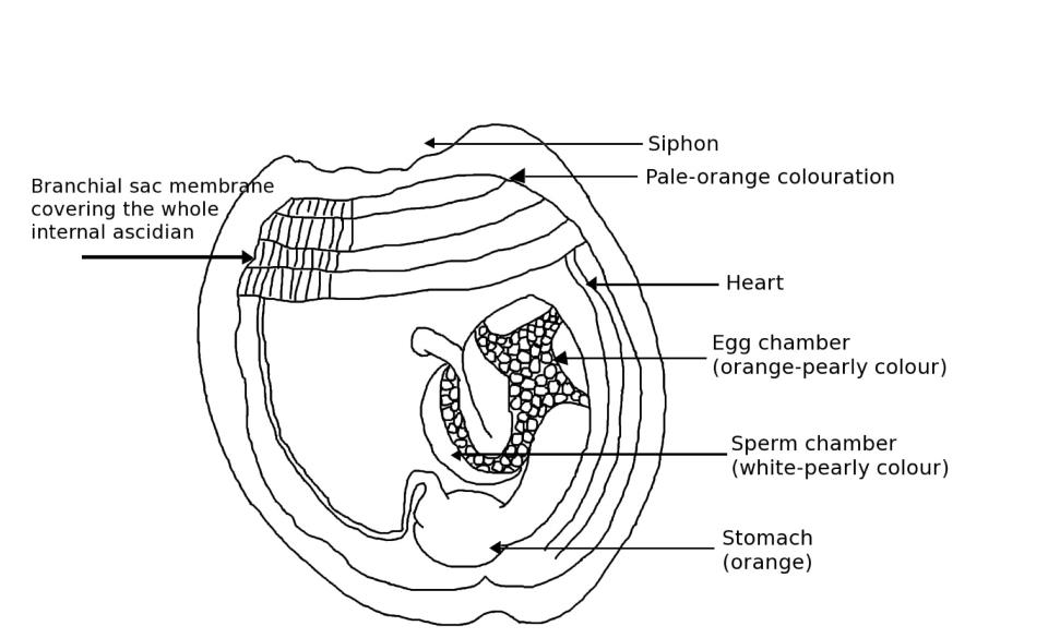
Anatomy
 A. glabra
 internal anatomy with colour indications.
Drawing by Emmanuelle Zoccola from personal observations and from Matther & Bennett (1993)
Digestive Track
In ascidians, the digestive track is disposed on a frontal plan instead than on a sagittal plan (Brien et al., 1948). The gut in separated into a foregut, a mid gut and a hind gut. First, food is filtered through the oral siphon that opens into the pharynx and the ciliated oesophagus. The pharynx includes the glandular endostyle producing mucus, which allows the coating of food particles and facilitates their movement along the digestive track (Krauss, 1978). The oesophagus then opens into the stomach, which presents four folds and a continuation of the oesophagus cilia (Brien et al., 1948). In the mid gut, the intestines,which are included in the primary loop of the gut (mid gut), are particularly long and are curved dorsally (Kott, 1985). Eventually, the rectum opens on the atrial siphon by the anus, which is slightly shifted to the left of the mid-dorsal axis (Brien et al., 1948).
Circulatory System
Hemal system of ascidians is well developed and the blood follows a define circuit (Ruppert et al., 2004). The blood contains several cell types. The lymphocytes form the connective tissues (totipotent cells) and differentiate in two types of amebocytes: the phagocytes (digesting and degrading alien cells) and the vacuolated or excretory cells containing the pigments (Brien et al., 1948; Ruppert et al., 2004). In A. glabra, the pigments are red hemoglobins (Kott, 1985).
The fluid-filled pericardial cavity consists in a hollow tube located ventrally to the gut loop (Ruppert et al., 2004). The pericardial cavity is opened at each end and related to the endostyle on one side and to the dorsal part of the pharynx on the other side (Brien et al., 1948; Ruppert et al., 2004). The heart is enclosed in the pericardial cavity, on the ventral part of the abdomen (Brien et al., 1948). Interestingly, the myocardium in ascidians is driven by waves and peristaltic contractions from one side of the heart to another, in one direction or the other (Brien et al., 1948; Ruppert et al., 2004). The blood circulation direction alternates generally every three or four minutes (Ruppert et al., 2004).
Muscles
Ascidia glabra muscles are irregularly distributed between the right and the left part of the body. There is an irregular mesh of muscles in the center of the right side of the body that straightens to form a border of parallel bands around the tunic border (Kott, 1985). However, on the left side of the body, there are only longitudinal muscles radiating from the branchial siphon in the anterior part of the body and there is no musculature over the gut at all (Kott, 1985). The muscles located on the right side are thus entirely responsible for the gut musculature.
Genital Organs
A. glabra, like most ascidians, are hermaphroditic (with both sexes). However, the egg sac and the sperm sac are distinct in A. glabra, although very closely located (Brien et al., 1948; Krauss, 1978). Indeed, both genital organs are located on the primary gut loop (Kott, 1985). This location allow the gametes to be released through the exhaling siphon during spawning events (Krauss, 1978). These event are usually synchronised to maximize fertilization chances (Krauss, 1978).
From left to right: microscopic observation of A. glabra oocytes at different development stages - A. glabra oocytes in situ - microscopic observation of A. glabra sperm
Photo by Emmanuelle Zoccola
Central Nervous System
Ascidian nervous system and sensory organs are poorly developed, probably due to their sedentary mode of life (Krauss, 1978). In adults, the CNS consists in a long neural ganglion, located between the two siphons, and some nerves extending from this ganglion (Krauss, 1978). Adults are also totally lacking any large sensory organ even if their body wall possesses individual chemoreceptor, photoreceptors and baroreceptors (Ruppert et al., 2004). Larvae, however, possesses an obvious notochord and sensory receptors such as chemoreceptors and photoreceptors to help them localize appropriate settlement sites (Krauss, 1978). |