External Morphology
Chitons are unsegmented, dorso-ventrally flattened and bilaterally symmetric. Cryptoplax larvaeformis is adapted to its cryptic life within crevices and as a result are stretched with its calcareous shell plates mostly separated. This form is called vermiform, they have a reduced foot and resemble a fat worm (Schwabe et al 2010).
Dorsal view:
The thick marginal tissue that surrounds the Chiton is called the girdle. The girdle visible when looking down on the Chiton is called the perinotum. The perinotum is covered by calcareous or corneous spines, spicules, scales, needles and/or hairs. Some or a combination of these structures may serve as sense organs, to feel, smell and/or detect chemicals in their surrounding environment (Schwabe et al 2010).

Figure 1: close up view of the anterior end of a Cryptoplax larvaeformis hight-lighted are some different aspects of the perinotum.
Calcareous shell plates:
  
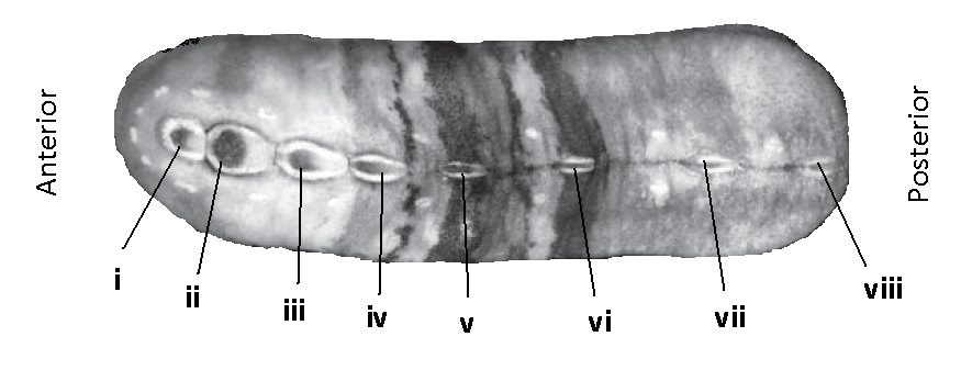
Figure 2: The dorsal view of a Cryptoplax larvaeformis, labeled are the calcareous shell plates. Picture adapted from Schwabe et al (2010).
In the Genus Cryptoplax most shell plates (or valves) separated, they are counted in an anterior to posterior direction and represented by Roman numerals (Figure 1). The fist shell plate (i) is also called the head plate, and the last plate (viii) is also called the tail plate, plates ii - vii are also known as intermediate plates
Aesthetes:
What are aesthetes?
Photoreceptory (light sensing) cells embedded in the shell plates (Blumrich 1891). There are two types Megalaesthetes and micraesthetes, as suggested by their names they are of differing sizes, small and big (Boyle 1972). They have been observed to take many different shapes and sizes which have little importance to what they can do, but rather represent inherent differences in shell growth (Currie 1992).
Where are they?
Embedded in the shell plates (Boyle 1972). They are exposed the environment via pores called micropores and macropores, and yes, they correspond to the two different sized aesthetes. These pores open up to an underlying complex canal system of cavities which are filled with sensory tissue, connecting the aesthetes to the nervous system (Schwabe 2010).
What are they not?
The common misconception: They are NOT eyes. They are simply pigmented cells that can detect light and send messages to the brain about where that light is coming from (Schwabe 2010, Currie 1992, Blumrich 1891).
Ventral view:
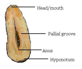
Chitons have a large fleshy foot and a separated head which is where you will find a centrally located mouth. At the other end of the foot (the posterior end) you can find a centrally positioned anus. The foot and the anus are surrounded by the mantle cavity also known as the pallial groove, here you can find the gills (ctenidia). The ventral side of the girdle is called the hyponotum. (figure 3)

Figure 3: The ventral view of a Cryptoplax larvaeformis.
|