Internal Anatomy
Form
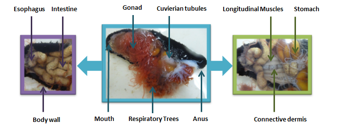
Holothuriums have a thick body wall, mainly composed of connective tissue dermis (Motokawa 1984). The body wall consists of three layers, a thin cuticle, a non-ciliated epidermis and a thick dermis (Ruppert, Fox & Barnes 2004). This dermis is comprised of ossicles made from calcarious pieces, which forms the ‘skeleton’ of the animal and help protect and toughen the body wall (Soltani et al. 2010). Generally, the body of H. leucospilota is pliant, due to adaptation to calmer waters where this species is located. Mechanical properties of the body wall display visco-elastic abilities, which give the species the ability to vastly deform itself. This is due to molecules in the body wall, which can quickly slip to rearrange themselves in a short time (Motokawa 1984). H. leucospilotacan also manipulate the overall tone of their body by employing the ‘stiffness-adjustable’ connective tissue, as seen in all echinoderms (Ruegg, 1971).
Movement
As a holothurian, H. leucospilota can be characterised by their 5 pairs of longitudinal muscles. These run from the mouth where they are attached near the peripharyngeal calcareous ring, to the posterior areas, near the cloaca (Conand, Morel & Mussard 1997). Similary to other soft bodies animals such as worms and sea anemone, the sea cucumber uses it long cyclindrical body, muscles, flexible body wall and fluid filled hydrostat to slowly move along the sandy substrate (Ruppert, Fox & Barnes 2004).
Feeding and Digestion
The pharynx opens into shortened esophagus, continuing to the stomach then a long, endodermal intestine, before reaching the anus (Ruppert, Fox & Barnes 2004). Mucus is used to help transport food to the stomach. The flow through gut of H. leucospilota, consists of a thin membrane intestine, often filled with sand, dead coral and any other rock debris picked up during non-selective deposit feeding (Conand, Morel & Mussard 1997). The intestine is coiled and fills a large section of the body and is divided into three loops, with the second loop being responsible for intestinal absorption via endocytosis and intracellular digestion. Intestines of H. leucospilota have been shown to lengthen on average to nearly one metre (Conand, Morel & Mussard 1997).
Respiration and Circulatory Systems
Respiration organs consist of two respiratory trees, consisting of a trunk with branches, with one each located on the left and right side of the body respectively. Respiration assists in the exchanges and elimination of metabolic wastes and involves the method of passively diffusing water. This also involved the water vascular system, which assists in the movement of sea water. Muscular pumping of the cloaca results in an intake of water via the anus, flooding the respiratory trees. The respiratory trees then contract to expell the water from the system. This allows the main source of gas exchange and the development of large body size for the species.
Although they do not have a heart, holothuroids have the most well developed hemal system out of all echinoderms (Ruppert, Fox & Barnes 2004). The well developed hemal system and ceolomic cavities is used in internal transport, including the water vascular system (Ruppert, Fox & Barnes 2004).
Reproduction
Individuals of H. leucospilota possess a single tuft of branching gonadal tubules (Purwati & Luong-van 2003). Gonadal tubules are attached by the gonad base near the mouth and hang freely in the body cavity from a transparent basis on the anterior end of the intestine (Conand, Morel & Mussard 1997., Purwati & Luong-van 2003). From this emerges a simple gonoduct, ending near the oral end of the individual (Purwati & Luong-van 2003). Males and female tubules differ in colour, with male tubules being creamy, white. Female tubules are more transparent, though change to a bright, reddish-orange colour with development of fecund ovaries (Purwati & Luong-van 2003).
Defence
When threatened, H. leucospilota discharges Cuvierian tubules from its anus, which are located internally at the base of the respiratory trees (Conand, Morel & Mussard 1997). These tubes are very sticky and it is believed their purpose is to entangle potential predators. Only a selection of the tubules are ejected at once and these are soon replaced by new Cuverian tubules (Nagabhushanam, Kumar & Sarojini 1994). These tubules can also be toxic to some other animals, thereby acting as a defence mechanism.
 |
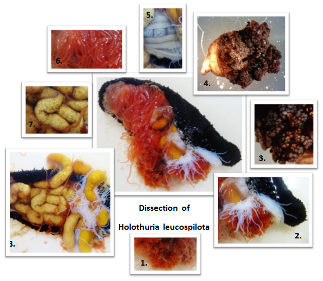
Figure 2. Dissection of H. leucsopilota, showing the internal anatomy.
|

|
1. Red respiratory trees
2. White cuvierian tubules
3. Close up of buccal podia
4. Single feeding tentacle
5. Longitudinal muscles
6. Gonad
7. Sediment in the intestine
8. Coiled intestine
|
|